Latest News
Highlight · Nov 19, 2022
Cornell University
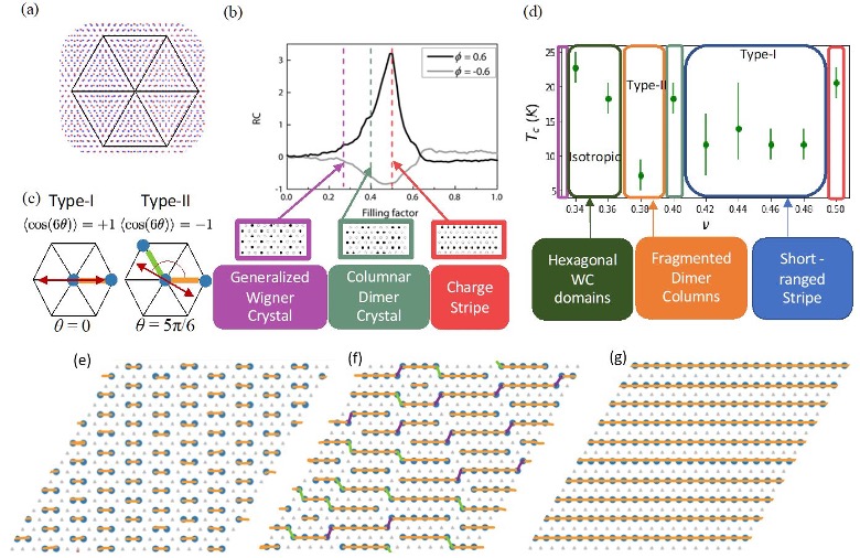
The Electronic Phase Diagram of Moiré Superlattices
Eun-Ah Kim (Cornell University)
Members of PARADIM’s in-house research team have used Monte Carlo simulations to study intermediate phases occurring at fractional electron counts between the charge-ordered states in the strong coupling limit. The team found two distinct stripe solid states to be each preceded by distinct types of nematic states and describes a microscopic mechanism that stabilizes each of the nematic states.
Highlight · Nov 18, 2022
University of California, Santa Barbara
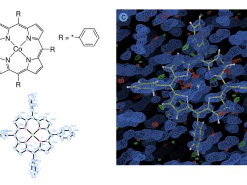
New first-of-its-kind Electron Diffraction Tomography (MicroED) microscope available at BioPACIFIC MIP facilities
BioPACIFIC MIP at UCLA
The BioPACIFIC MIP team has completed the construction and installation of a state-of-the-art transmission electron microscope (TEM) and has made the instrument available to users.
News · Nov 18, 2022
University of California, Santa Barbara
BioPACIFIC MIP has signed off on the state of the art microED
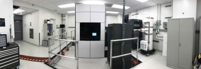
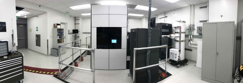
UCLA has introduced a shared-use electron microscope, the ThermoFisher Scientific Spectra 300C, for studying atomic structures of bio-sourced compounds. This tool enables rapid structure determination through micro-electron diffraction, significantly reducing analysis time. It supports the BioPACIFIC MIP collaboration aimed at developing sustainable bio-based materials, benefiting researchers at UCLA, UCSB, and beyond.
News · Nov 18, 2022
BioPACIFIC MIP has signed off on the state of the art microED
There’s a new shared-use electron microscope at UCLA for investigators seeking to elucidate the atomic structures of bio-sourced monomers and small molecules. The NSF’s BioPACIFIC MIP now offers access to its ThermoFisher Scientific Spectra 300C transmission electron microscope (TEM) housed at the California NanoSystems Institute at UCLA.
Highlight · Nov 14, 2022
Cornell University
From Machine Learning to Discovery of New Family Member
Lena F. Kourkoutis and Darrell G. Schlom (Cornell University)
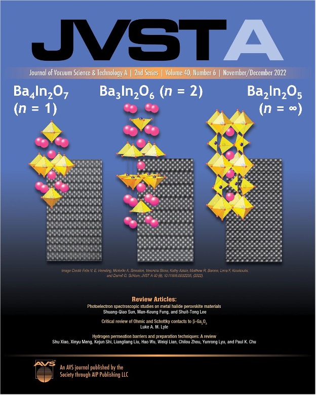
Thin films of Ba3In2O6 had never been made and PARADIM researchers were able to grow the desired phase using a new variant of molecular-beam epitaxy (MBE), suboxide MBE, developed in PARADIM. In addition to succeeding in the growth of the targeted phase, Ba3In2O6, PARADIM researchers also discovered a new barium indate phase with an even simpler crystal structure, Ba4In2O7, that is analogous to the first high-temperature cuprate superconductor discovered, (La,Ba)2CuO4.
News · Nov 1, 2022
Cornell University
PARADIM Researcher Berit Goodge Awarded Third Prize in APL Materials Excellence in Research Award
By PARADIM News
Berit Goodge received third prize in the APL Materials Excellence in Research Award for her study on lattice disorder's impact on superconductivity in Sr₂RuO₄.
Highlight · Oct 31, 2022
Cornell University
Lorentz Ptychography for high-resolution, high sensitivity magnetic imaging
David A. Muller (Cornell University)
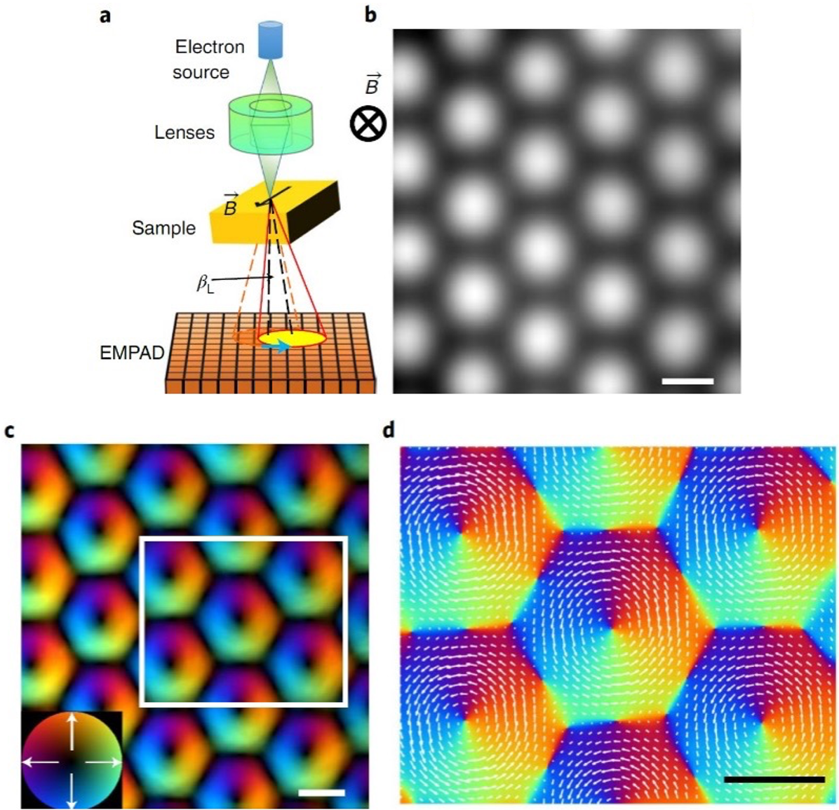
The study establishes a quantitative, high-resolution magnetic microscopy technique that can reveal nanoscale spin textures, especially magnetization discontinuities and topological defects in nanomagnets. The technique’s high-dose efficiency should also make it well suited for the exploration of magnetic textures in electron radiation-sensitive materials such as organic or molecular magnets.
Highlight · Oct 14, 2022
University of California, Santa Barbara
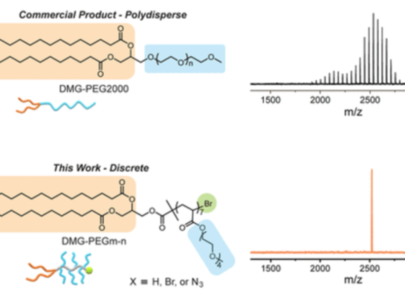
Discrete Libraries of Amphiphilic Poly(ethylene glycol) Graft Copolymers: Synthesis, Assembly, and Bioactivity
Junfeng Chen, Aoon Rizvi, Joseph P. Patterson, and Craig J. Hawker (UCSB and UCI)
Demonstration of a novel strategy for the scalable synthesis of multiple libraries of discrete lipid poly(ethylene glycol) (DMG-PEG) derivatives, with precise control over the structure and molecular weight.
Highlight · Oct 3, 2022
University of California, Santa Barbara
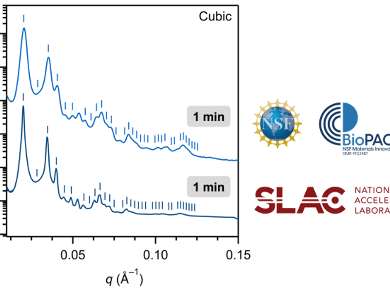
Efficient Creation and Morphological Analysis of ABC Triblock Terpolymer Libraries
Craig Hawker and Christopher Bates (UCSB)
A versatile and scalable strategy to separate parent triblock terpolymers into libraries of fractionated samples spanning a wide range of compositions was developed.
News · Oct 1, 2022
Cornell University
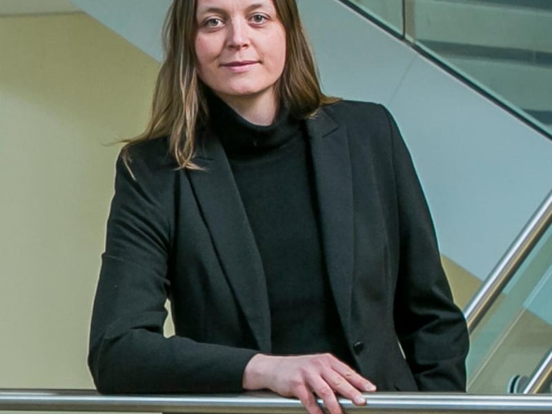
Lena Kourkoutis Named 2022 APS Fellow for Advances in Electron Microscopy
By PARADIM News
Lena Kourkoutis was elected a 2022 Fellow of the American Physical Society for her groundbreaking work in cryogenic scanning transmission electron microscopy.
Showing 151 to 160 of 363